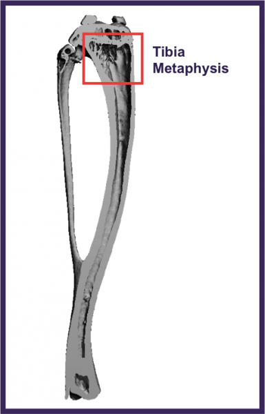
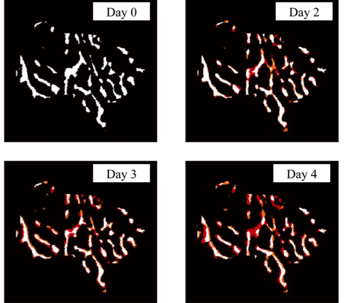
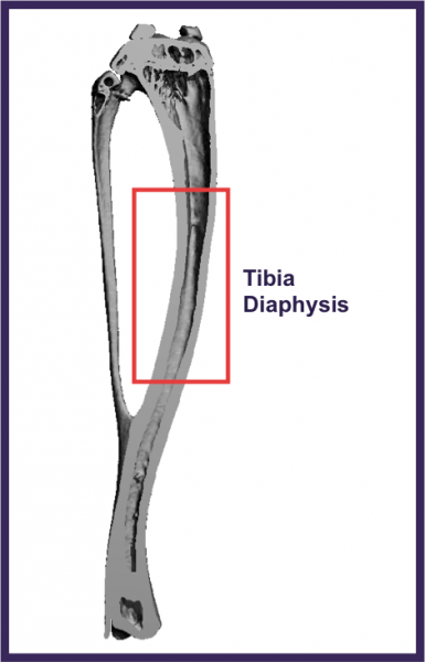
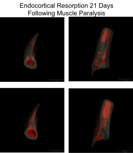
The goal of this study is to track the onset and migration of bone loss induced by transient muscle paralysis with the hopes of using this data to elucidate the drivers of osteoclastic bone resorption in this model. Bone loss induced by transient muscle paralysis is an osteoclast driven process that results in dramatic endocortical resorption at the tibia midshaft, while leaving the periosteal surface relatively unchanged. This unchanged periosteal surface can be used as a registration landmark to superimposed serial micro-CT images as a way to track the spatial and time dependent resorption that occurs on the endocortical surface. 3-Dimensional images of bone loss seen below, highlight the complex, yet highly reproducible spatial pattern of cortical bone loss observed in our model. Expansion of our image registration technique has allowed for tracking of focal bone loss initation in the highly metabolically active trabecular bone adjacent to the growth plate (bottom).
Endocortical Resorption Following Muscle Paralysis Occurs in a Complex, Yet Spatially Consistant Heterogenious Pattern
Initation of Osteoclastic Resorption Following Muscle Paralysis Occurs Acutely in Adjacent to the Proximal Growth Plate
