Surgery for Atraumatic Instability of the Shoulder

About surgery for atraumatic instability

This is the information which might be shared with patients as they consider surgical management of atraumatic instability. Before it can be applied to a specific clinical situation, however, it needs to be tailored to the patient, the problem, and the surgeon.

Management of atraumatic instability

A shoulder may have problems with instability even though there has not been a major traumatic injury. As a result, the shoulder may slip or feel unstable with certain activities. Most often, atraumatic shoulder instability can be managed by restoring the normal strength and coordinated use of the shoulder through a reconditioning program. Rarely, the instability is so severe that surgery is considered as an adjunct to the exercise and training program. Shoulder surgery itself cannot "fix" the problem of atraumatic instability because there is no simple rip to sew up. Instead, the goal of shoulder surgery for this type of instability is to tighten the tissues around the shoulder joint, restricting its range of motion but also helping add some stability.

Details of the surgery

Careful consideration of indications for surgery

The ability of surgery alone to cure atraumatic instability is limited. Usually there is no single lesion that can be repaired. Most of the factors providing midrange stability cannot be enhanced by surgical reconstruction. Problems of poor neuromuscular control or relative glenoid flatness do not have surgical solutions. Even after a snug capsulorrhaphy, the midrange stabilizing mechanisms of balance and concavity compression must be optimized through muscle strengthening and kinematic training. Otherwise, excessive loads will be applied to the surgically tightened glenohumeral capsule, leading to stretching and failure of the surgical reconstruction.

In this light, the indications for surgical treatment of atraumatic instability need to be carefully considered. First, the patient must have major functional problems that are clearly related to atraumatic glenohumeral instability. Second, the patient must clearly understand that good strength and kinematic technique are the primary stabilizing factors for the shoulder rather than capsular tightness. Third, the patient must have participated in a strengthening and training program conscientiously and recognize that strength and proper technique will continue to be major stabilizing factors for the shoulder even after reconstructive surgery is performed. The patient must also recognize that capsulorrhaphy is designed to stiffen the shoulder: the surgery will compromise the range of motion in the hope of gaining stability. If attempts to regain normal range are made early on, instability is likely to recur. Thus the limitations imposed by surgical capsulorrhaphy may be incompatible with the goals of normal or supernormal range of motion. Therefore, gymnasts, dancers, and baseball pitchers may not be good candidates for this surgical procedure. Similarly, this procedure has a limited ability to hold up under the demands of heavy physical labor unless it is accompanied by a superb strength and kinematic rehabilitation program. Finally, the patient must understand that rehabilitation after this procedure is protracted. It is important that the shoulder be immobilized in a brace for a month, during which time muscles get weak and normal kinematics are lost. After this month of immobilization many months are required for the reestablishment of good strength and shoulder kinematics. In spite of the best operative and postoperative management, the success of this procedure in reestablishing normal shoulder function is substantially less than procedures for traumatic instability.

The foregoing is a large amount of very important information that must be understood by the patient considering the surgical procedure. The situation is further complicated by the fact that many patients who present with atraumatic midrange instability are young and may have difficulty understanding and accepting the ramifications of this information. Thus, during the preoperative discussions with young patients it may be important that parents participate actively. We find that many families who present requesting that "the shoulder be fixed" are prepared to work more diligently on the non-operative program after this discussion. We provide detailed information to patients and families interested in the surgical management of atraumatic multidirectional instability.

Surgical technique

The essential elements of the procedure are reconstruction of the rotator interval capsule/coracohumeral ligament mechanism and reduction of the posteroinferior capsular recess. These goals can be accomplished only through an anterior surgical approach. Thus, we usually approach a repair for atraumatic instability from the front, even if the predominant direction of instability appears to be posterior. There are additional advantages of the anterior approach. It is cosmetically superior to the posterior approach. It is accomplished without incising the critical external rotator cuff musculature. Finally, when the capsule is advanced anterosuperiorly on the humeral side, elevation of the arm anteriorly results in additional tightening of the inferior and posterior capsule.

The shoulder is approached through a low anterior axillary incision, entering the deltopectoral groove medial to the cephalic vein. The clavipectoral fascia is divided up to the level of the coracoacromial ligament. The axillary nerve is palpated medially as it courses across the subscapularis and passes inferiorly toward the quadrangular space. The superior edge of the subscapularis is then identified by palpating the rotator interval lateral to the coracoid process and medial to the bicipital groove. The triad of anterior humeral circumflex vessels mark the inferior border of the subscapularis. The subscapularis tendon is sharply and carefully dissected from the subjacent capsule.

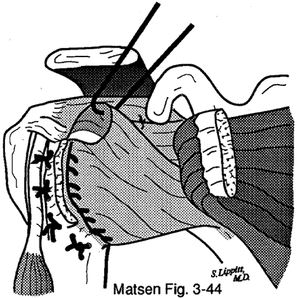
A substantial defect in the rotator interval is seen consistently in the AMBRII syndrome. This defect is bordered by the capsule adjacent to the supraspinatus overlying the biceps tendon superiorly, the anterior capsule and subscapularis anteroinferiorly, the coracoid process, and the transverse humeral ligament. This defect is accentuated by pushing the humeral head posteriorly. Sutures of number 2 non-absorbable material are securely placed in the superior edge of the defect and then passed across to the inferior edge of the defect. When these sutures are tied, a strong rotator interval capsule is reconstructed.

The anterior capsule is incised from the humeral neck beginning just below the top of the lesser tuberosity. Traction sutures are placed in the capsule. With the axillary nerve protected and with the arm in adduction and neutral rotation, the anterior inferior and the inferior capsule are incised from the humeral neck. This dissection is continued until superiorly directed traction on the capsular flap causes the capsule to tighten on a finger placed in the posteroinferior capsular recess. Usually this point is reached when the capsule is released just past the inferior (6 o'clock position) on the humeral neck, sectioning the posterior band of the inferior glenohumeral ligament.

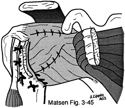
After the capsular release, a bony trough is created in the anteroinferior humeral neck adjacent to the articular surface using a power burr. Holes are made in the humeral neck lateral to the groove and sutures are passed through these holes into the groove for reattachment of the capsule securely to bone. With the arm at the side and in neutral rotation and with strong anterior superior traction on the sutures to obliterate the posterior inferior recess, the sutures from the groove are passed through the lateral edge of the capsule. Tying these sutures securely fixes the capsule in its advanced position. This step needs to be accomplished with excellent direct vision to be sure the bites in the capsule are sufficiently inferior to tighten it to the groove and to assure the safety of the axillary nerve. The surgeon must assure that pulling up on these sutures obliterates the posterior inferior recess. If this is not the case, either the inferior capsular release was insufficient or the sutures were not placed sufficiently inferior.

This repair to the groove is continued anteriorly up the humeral neck.

Redundant anterior superior capsule is folded down over the previous repair to reinforce it.

At this point the shoulder is checked to ensure that internal rotation of the abducted arm is limited to 45 degrees below the horizontal, that the posterior drawer is less than 50 percent of the humeral head diameter and that external rotation of the arm at the side is 30 degrees. Excessive internal rotation of the abducted arm indicates the inferior capsule was not advanced sufficiently. Excessive translation on the sulcus test indicates that the rotator interval capsule was insufficiently tightened. Excessive limitation of external rotation indicates that the anterior capsule was tightened too much.

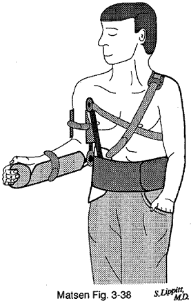
The subscapularis is then repaired to its normal anatomic insertion. After a standard wound closure, the arm is placed in a prefitted "handshake" orthosis with the arm in neutral rotation and slight abduction.

 
Click to enlarge
Figure 1 - Essential elements of the technique
 
Click to enlarge
Figure 2 - Reconstruction of a strong rotator interval capsule
Click to enlarge
Figure 3 - Posteroinferior capsular recess
Click to enlarge
Figure 4 - Power burr creating a bony trough in the anteroinferior humeral neck adjacent to the articular surface
Click to enlarge
Figure 5 - The sutures from the groove are passed through the lateral edge of the capsule
Click to enlarge
Figure 6 - Fixing the capsule in its advanced position
Click to enlarge
Figure 7 - Folding the redundant anterior superior capsule over the previous repair to reinforce it
Click to enlarge
Figure 8 - "Handshake" orthosis

Rehabilitation

Postoperative rehab

With the arm in the orthosis, the patient is started on grip strengthening, elbow range of motion, isometric external rotation, and isometric abduction shoulder exercises. The brace is usually continued for one month, although longer periods may be used for individuals who are extremely lax, and shorter periods may be used for individuals over 25 years of age who may be prone to excessive stiffness.

The patient is then weaned from the orthosis over a period of a week. During this time the patient is taught to elevate the arm in the coronal plane only, to continue the cuff and deltoid strengthening, and to avoid any activities that may challenge the repair. From this point, range of motion is gained only with active exercises; no passive stretching is used. Lifting of more than 10 pounds is delayed for six months. Sports are delayed for at least one year after surgery and are permitted only if the patient has excellent strength and dynamic control of the shoulder.

The patient needs to understand in detail the importance of this program.

Rehabilitation instructions

These are typical instructions given to a patient after a repair for atraumatic instability. However, the program will vary with the surgeon, the patient, and the repair. If you have had such a repair, only your surgeon can give you instructions on the postoperative management. Do not do any exercises after surgery except after consultation with your physician.

The arm may be placed in a brace to assure that it heals properly. If this brace is not comfortable, be sure to let your doctor know immediately. For one month after the operation your arm must stay in the brace. If someone can hold your arm in the correct position for you during bathing, you may briefly remove it for this purpose. Otherwise, you should clean yourself twice a day using an alcohol sponge underneath the straps of the brace. You can put on a shirt if someone can help you by keeping your arm in the correct position while your arm is threaded through the sleeve. Otherwise you should wear loose-fitting clothes over the brace.

Each day you can loosen the forearm straps to put your elbow through a range of motion.

Your doctor may start exercises such as these while you are still in your brace. First, you need to maintain the strength of your grip by squeezing a ball, sponge, or putty several hundred times a day. Second, you should perform 3 minutes of gentle isometric exercises at least three times a day against some fixed object--pushing your wrist outward, pushing your hand forward, pushing your elbow outward, and pushing the elbow back. These exercises are designed to maintain your muscle tone. The shoulder is not moved during these exercises. The exercises should be comfortable.

You should return to the office at one month after surgery. If everything is healing properly, you may begin weaning yourself from the brace at that time.

In the weaning process you will start moving your shoulder out to the side, avoiding the positions that used to be symptomatic for you. You may find it reassuring to sleep in your brace for another week. You will regain your motion on your own, moving your shoulder under its own motor power and specifically avoiding any stretching. You need to continue your previous isometric exercises and can add progressively the same exercises you did before surgery.

Always avoid "checking" your shoulder to see if it is stable. You must not lift more than 10 pounds for the first six months after your surgery.

After four months you can begin gentle, well-controlled, repetitive activities with your shoulder, such as swimming or using a rowing machine, provided that these activities are comfortable for you.

You cannot return to contact sports or heavy work for at least one year after this surgery, and then only if you have excellent strength and coordinated control of your shoulder.

If you have any questions at any time, please let your doctor know.