Last updated: Monday, February 4, 2013
The clinical manifestations of the various clinical forms of cuff disease include difficulties with shoulder stiffness weakness instability and roughness. (Matsen Lippitt 1994 Samilson and Binder 1975)
Stiffness limits passive range of motion and frequently causes pain at the end point of motion as well as difficulty sleeping.
Stiffness as limitation
Stiffness is most common in partial thickness cuff lesions but may also be associated with full thickness cuff defects. (Jackson 1976) Stiffness may be demonstrable as limitations of:
- internal rotation with the arm in abduction (degrees from the neutral position) (see figure 1)
- reach up the back (posterior segment reached with the thumb) (see figure 2)
- cross-body adduction (centimeters from the ipsilateral antecubital fossa to the contralateral acromion or coracoid) (see figure 3)
- flexion (degrees from the neutral position) (see figure 4) or
- external rotation (degrees from the neutral position) (see figure 5).
Weakness or pain on muscle contraction limits the function of the shoulder with cuff disease.
More about weakness
Tendon fibers weakened by degeneration may fail without clinical manifestations or may produce only transient symptoms interpreted as "bursitis" or "tendinitis." A greater injury is required to tear the cuff of individuals at the younger end of the age distribution. Traumatic glenohumeral dislocations in individuals over the age of 40 have a strong association with rotator cuff tears. These traumatic cuff tears commonly involve the subscapularis producing weakness in internal rotation. Neviaser et al (Neviaser Neviaser 1993) reported on thirty-seven patients older than 40 years of age in whom the diagnosis of cuff rupture was initially missed after an anterior dislocation of the shoulder. The weakness from the cuff rupture was often erroneously attributed to axillary neuropathy. Eleven of these patients developed recurrent anterior instability that was due to rupture of the subscapularis and anterior capsule from the lesser tuberosity. None of these shoulders had a Bankart lesion. Repair of the capsule and subscapularis restored stability in all of the patients with recurrence.
Sonnabend reported a series of primary shoulder dislocations in patients (Sonnabend 1994) older than 40 years of age. Of the 13 patients who had complaints of weakness or pain after 3 weeks eleven had rotator cuff tears. Toolanen found sonographic evidence of rotator cuff lesions in 24 of 63 patients over the age of 40 years at the time of anterior glenohumeral dislocation. (Toolanen Hildingsson 1993)
Manifesting weakness
Even though patients with full thickness cuff defects may still retain the ability to actively abduct the arm (Neviaser 1971) significant tendon fiber failure is usually manifest by weakness on manual muscle testing. (Brems 1987 Jan Hawkins Misamore 1985 Leroux Codine 1994 Leroux Thomas 1995) Isometric testing of muscle strength prevents confusion with symptoms which may arise from shoulder movement (such as those associated with subacromial abrasion). While the individual cuff muscles cannot be specifically isolated the following isometric tests are reasonably selective (see figure 6):
- supraspinatus: isometric elevation of the arm held in 90 of elevation in the plane of the scapula and in mild internal rotation.
- subscapularis: isometric internal rotation of the arm with the elbow flexed to 90 and the hand held posteriorly just off the waist.
- infraspinatus: isometric external rotation of the arm held at the side in neutral rotation with the elbow flexed to 90.
These simple manual tests are helpful in characterizing the size of the tendon defects from single tendon tears involving only the supraspinatus to two tendon tears involving the supra and infraspinatus to three tendon tears involving the subscapularis as well.
Individuals with partial thickness cuff lesions have substantially more pain on resisted muscle action than those with full thickness lesions. This phenomenon is analogous to the observation that partial tears of the Achilles tendon partial tears of the patellar tendon and partial tears of the origin of the extensor carpi radialis brevis are more painful on muscle contraction than when the complete structure is ruptured or surgically released. Fukuda and coworkers (Fukuda Mikasa 1987) characterized patients with partial-thickness cuff tears as having pain on motion crepitus and stiffness. They observed that patients with bursal side tears seemed more symptomatic than those with deeper tears due to the resulting problems with roughness of the articulation between the upper surface cuff and the under surface of the coracoacromial arch.
Some have suggested that weakness from pain inhibition can be distinguished from weakness from tendon defect by a subacromial injection of local anesthetic. (Ben-Yishay Zuckerman 1994 Lindblom and Palmer 1939) If cuff dysfunction has been present for more than a month or so it may be accompanied by supraspinatus and infraspinatus muscle atrophy. Subtle atrophy can be seen most easily by casting a shadow from a light over the head of the patient.
Defects in the cuff
As pointed out by Codman (Codman 1934b) defects in the cuff can often be palpated by rotating the proximal humerus under the examiner's finger placed at the anterior corner of the acromion. The perimeters of the "divot" left by a defect in the supraspinatus are particularly easy to palpate. The defect is usually just posterior to the bicipital groove and medial to the greater tuberosity.
The inability to keep the head centered in the glenoid may result from cuff disease.
Acute tears
Acute tears of the subscapularis may contribute to recurrent anterior instability. (Neviaser Neviaser 1993 Sonnabend 1994 Toolanen Hildingsson 1993)
Chronic loss of compressive effect
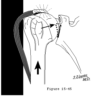
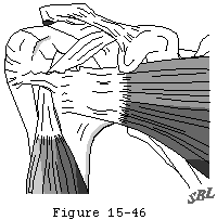
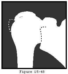
Chronic loss of the normal compressive effect of the cuff mechanism and of the stabilizing effect of the superior cuff tendon interposed between the humeral head and the coracoacromial arch may contribute to superior glenohumeral instability. (Flatow Raimondo 1996 Flatow Soslowsky 1994 Lazarus Harryman II 1995 February 16-21 Poppen and Walker 1976 Ziegler Matsen III 1996) Superior instability is magnified in the presence of wear of the upper glenoid rim (see figures 7-9) (Neer Craig 1983) and when the normal supportive function of the coracoacromial arch is lost from erosion or surgical removal. (Wiley 1991)
Roughness associated with cuff disease manifests itself as symptomatic crepitus on passive glenohumeral motion.
More about roughness
Bursal hypertrophy secondary changes in the undersurface of the coracoacromial arch loss of the integrity of the upper aspect of the cuff tendons degenerative changes of the tuberosities may all contribute to subacromial abrasion. Crepitus from subacromial abrasion is easily detected by placing the examiner's thumb and fingers on the anterior and posterior aspects of the acromion while the humerus is moved relative to the scapula (see figures 10 and 11). In that many shoulders demonstrate asymptomatic subacromial crepitus it is important during the examination to ask whether the crepitus noted by the examiner is directly related to the patient's complaints.
Rotator cuff tear arthropathy is another cause of roughness associated with cuff disease. This term coined by Neer and coworkers (Neer Craig 1983) denotes the loss of the glenohumeral articular surface in association with a massive rotator cuff deficiency (see figure 12). These authors described 26 shoulders of which over 75% were in female patients. The average age was 69 years; 20 per cent had evidence of contralateral cuff arthropathy and 75 per cent had no history of trauma. Typically the shoulders were swollen the muscles atrophic and the long head biceps ruptured; passive elevation was limited to an average of 90 degrees of elevation and 20 degrees of external rotation (a degree of limitation atypical of uncomplicated cuff tears). Often the shoulder demonstrated anteroposterior instability. Collapse of the proximal humeral subchondral bone was a common observation. Glenoid greater tuberosity acromial and lateral clavicular erosion were also commonly observed. The authors hypothesized that the arthropathy resulted from both mechanical factors (such as anteroposterior instability and superior migration of the humeral head) (see figures 13-17) and nutritional factors (such as loss of a closed joint space lack of normal diffusion of nutrients to the joint surface and disuse). To this list could be added the disruption of the tendinous--osseous circulation entering through the subscapular anterior humeral circumflex and suprascapular vessels. This condition is distinct from osteoarthritis rheumatoid arthritis avascular necrosis and neurogenic arthropathy. (Neer 1983)


