Last Updated: Monday, February 4, 2013
The humeral head and the glenoid normally articulate through smooth congruent and well lubricated joint surfaces.
About arthritis of the shoulder
Glenohumeral arthritis results when these joint surfaces are damaged by congenital metabolic traumatic degenerative vascular septic or nonseptic inflammatory factors. These conditions are common especially in older populations where the prevalence approaches 20% (Chard and Hazleman 1987; Jenkinson et al. 1989; van Schaardenburg et al. 1994) In degenerative joint disease the glenoid cartilage and subchondral bone are typically worn posteriorly sometimes leaving intact articular cartilage anteriorly. The cartilage of the humeral head is eroded in a pattern of central baldness often surrounded by a rim of remaining cartilage and osteophytes. In inflammatory arthritis the cartilage is usually destroyed evenly across the humeral and glenoid joint surfaces. Cuff tear arthropathy occurs when a chronic large rotator cuff defect subjects the uncovered humeral articular cartilage to abrasion by the undersurface of the coracoacromial arch. The erosion of the humeral articular cartilage begins superiorly rather than centrally. Neurotrophic arthropathy arises in association with syringomyelia diabetes or other causes of joint denervation. The joint and subchondral bone are destroyed because of the loss of the trophic and protective effects of its nerve supply. In capsulorrhaphy arthropathy prior surgery for glenohumeral instability leads to joint surface destruction. In this situation excessive anterior or posterior capsular tightening forces the head of the humerus out of its normal concentric relationship with the glenoid fossa. The eccentric glenohumeral contact increases contact pressures and joint surface wear. Most commonly overtightening of the anterior capsule produces obligate posterior translation posterior glenoid wear and central wear of the humeral articular cartilage.
Mechanics of arthritis and arthroplasty
Four basic mechanical characteristics are essential to the function of the normal shoulder: motion stability strength and smoothness. Each of these is commonly compromised in the arthritic shoulder and can potentially be restored by shoulder arthroplasty. The approach to glenohumeral arthritis is guided by an understanding of the necessary elements for optimal shoulder mechanics.
The requisites of a normal range of glenohumeral motion include:
a. Normal capsular laxity
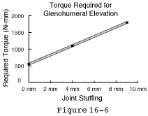
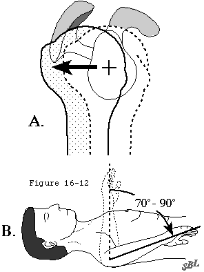
Normal capsular laxity allowing a full range of rotation. The glenohumeral capsule is normally lax through most of the functional range of shoulder motion. (Harryman et al. 1992; Lippitt et al. 1994) As the joint approaches the limit of its range the tension in the capsule and its ligaments increases sharply serving to check the range of rotation (see figure 1). In many conditions requiring shoulder arthroplasty the capsule and ligaments are contracted limiting the range of rotation. Shoulder arthroplasty tends to further tighten the capsule because a degenerated and collapsed humeral head is replaced by a relatively larger prosthesis and because a glenoid component is added to the surface of the glenoid bone which may consume more space than the degenerated cartilage it replaces (see figure 2) and "stuff" the joint. Unless sufficient capsular releases (see figures 3 and 4) have been performed to accommodate this stuffing the joint is "overstuffed." If the joint is overstuffed joint motion is limited (see figure 5) and more torque (muscle force) is required to move the arm (see figure 6).
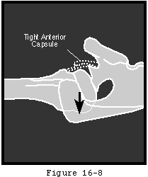
Harryman et al (Harryman et al. 1995) determined that all motions including flexion external and internal rotation and maximal elevation are diminished when a too-large humeral head prosthesis is implanted. Furthermore this overstuffing causes obligate translation of the head to occur on the glenoid; for example forced posterior translation occurs when external rotation is attempted against a tight anterior capsule (see figures 7 and 8). Thus if normal capsular laxity is lacking unwanted translation and eccentric glenoid loading may result.
In arthroplasty surgery the amount of stuffing can be estimated by adding the thickness of the glenoid component to the difference between the amount of intraarticular humerus replaced and the amount of humerus resected. To be comparable the measurement of the amount of humeral head resected and the measurement of the amount of intraarticular humeral prosthesis added must both be made from the cut surface of humeral neck to the articular surface. In modular humeral heads the amount of bone replaced must include the thickness of the collar and the exposed part of the Morse taper stem as well as the head itself see (see figure 2).
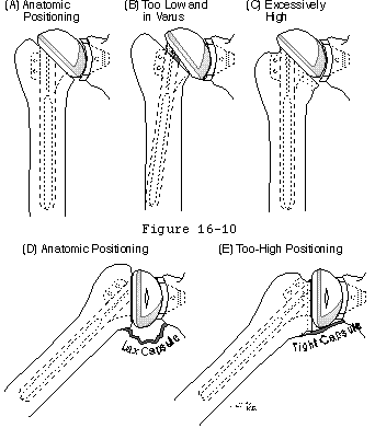
The amount of stuffing from the glenoid component is related primarily to its thickness as well as the amount of reaming the presence or absence of cement between the component and bone and the use of bone grafts. The thickness of currently available glenoid components varies from 3 to over 15 mm. Thicker glenoid polyethylene may help manage contact stresses and may have superior wear properties. (Friedman LaBerge et al. 1992) Metal-backed glenoid components affect load transfer and offer opportunities for screw fixation and tissue ingrowth. (Cofield and Daly 1992) However both thicker polyethylene and metal-backing contribute to joint stuffing which becomes particularly problematic in shoulders that remain tight even after soft tissue releases. Overstuffing may also predispose the reconstructed shoulder to instability. (Cofield and Daly 1992) The amount of stuffing from the humeral component is determined by both the geometry of the component and the position in which it is placed. The size of the intraarticular aspect of the humeral component is related to its design including its radius of curvature the percent of the sphere represented by its articular surface and the distance between the base of its collar and the articular surface of the prosthesis (see figures 2 and 9). The position of the component also has a major effect on the degree to which it stuffs the joint. A component inserted into varus will disproportionately stuff the joint when the arm is at the side; this outcome is more likely when the stem of the prosthesis does not fit the humeral canal snugly. A component inserted excessively high will tighten the capsule as the arm is elevated (similar to a mechanical cam) and limit the range of elevation (see figure 10).
Cadaver studies (Matsen Lippitt Sidles et al. 1994) indicate that less that 10 mm of overstuffing can reduce normal capsular laxity by as much as 50%. The overstuffed shoulder is predisposed to obligate translation.
The variables of component size and capsular release are under surgeon control. As a rules of thumb for judging capsular laxity at the time of surgery
- the humeral head should translate approximately 15 mm on the posterior drawer
- the abducted arm should allow 70 degrees of internal rotation and
b. Humeral articular surface area
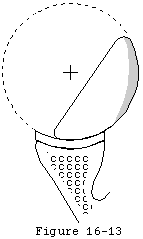
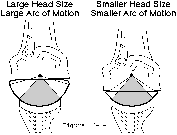
A substantial and properly located humeral articular surface area allowing a large unimpeded rotational range. Humeral articular surfaces that comprise only a small portion of the sphere (see figure 12) predispose to abutment of the rim of the glenoid against the tuberosities or anatomic neck of the humerus (see figure 13). (Ballmer Lippitt Romeo et al. 1994) The normal extent of the humeral joint surface can be restored with appropriate positioning of the appropriate prosthesis at the time of joint replacement (see figure 9).
c. Glenoid articular surface
A glenoid articular surface which comprises a relatively small portion of the sphere in comparison to that of the humerus. If the prosthetic glenoid joint surface area is large compared to that of the humerus abutment of the prosthesis against the humeral neck or tuberosities can restrict joint motion (see figures 13 and 14).
d. Absence of blocking osteophytes
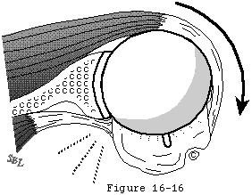
Osteophytes predispose to contact with the glenoid which can impair motion (see figure 15). Blocking osteophytes must be completely resected at the time of joint reconstruction (see figure 16).
e. Unrestricted humeroscapular motion interface
Normally 3 to 4 centimeters of excursion takes place at the upper aspect of this interface between the coracoid muscles and the subscapularis (see figure 17). Adhesions or "spot welds" between the proximal humerus and cuff on one hand and the deltoid and coracoacromial arch on the other can limit motion even if the intraarticular aspect of the arthroplasty is perfectly balanced. Lysing humeroscapular spot welds is an important early step in arthroplasty of the shoulder.
The requisites of glenohumeral stability include:
a. Humeral articular surface area
An anatomically oriented and sufficiently extensive humeral articular surface area. The orientation of the humeral articular surface can be described in terms of the humeral head center line; a line passing through the center of the humeral articular cartilage and through the center of the anatomic neck. This line usually makes a valgus angle of about 130 degrees with the humeral shaft. The humeral head center line usually makes a retroversion angle of about 30 degrees with the axis of elbow flexion. (Cofield 1984; Collins Harryman Lippitt et al. 1991; Figgie Inglis Goldberg et al. 1988; Friedman Thornhill Thomas et al. 1989; Hawkins Bell and Jallay 1989; Neer 1990; Neer and Kirby 1982; Pearl and Lippitt 1993; Roper Paterson and Day 1990; Weiss Adams Moore et al. 1990) Recent studies (Kronberg Brostrom and Soderlund 1990; Pearl and Volk 1995; Roberts Foley Swallow et al. 1991) point out that mean humeral retroversion varies widely from 7 to 50 degrees. Hernigou et al pointed out the importance of clearly defining the reference system when measuring humeral version. (Hernigou Duparc and Filali 1995)
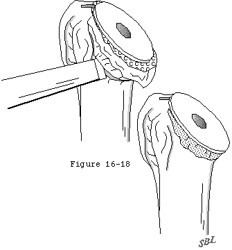
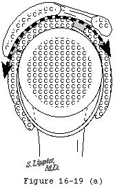
The extent of the humeral articular surface area is another critical determinant of stability. In the arthritic glenohumeral joint stability can be compromised by a reduced amount of available humeral articular surface. Similarly a prosthetic surface area that comprises only a small part of the total sphere (see figures 18 and 19) can predispose to instability in the same way as does a Hill-Sach's defect in traumatic instability by offering less contact area for joint surface contact (see figure 20).
b. An anatomically oriented glenoid
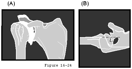
The glenoid center line the line perpendicular to the center of the glenoid fossa is usually relatively closely aligned with the plane of the scapula (see figures 21 and 22). In the arthritic glenohumeral joint stability may be compromised by abnormal glenoid version (see figures 23-25). Friedman et al (Friedman Hawthorne and Genez 1992) and Mullaji et al (Mullaji Beddow and Lamb 1994) have used CT to document that arthritic involvement may alter the glenoid version. The orientation of the glenoid prosthesis should be normalized as a part of the arthroplasty procedure (see figures 26-28).
c. Glenoid concavity
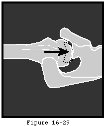
A glenoid concavity with sufficiently large effective arcs. The arc of the glenoid determines the maximal angles that the net humeral joint reaction force can make with the glenoid center line before dislocation occurs (see figure 29).
In the arthritic joint the effective glenoid arc can be diminished by wear or inflammation for example posterior wear is typical of glenohumeral osteoarthritis (see figures 24 and 30) and capsulorrhaphy arthropathy (see figure 31) while central erosion of the glenoid is typical of rheumatoid arthritis (see figure 32). At arthroplasty the effective glenoid arcs need to be restored (see figure 28).
d. Control of the net humeral joint reaction force
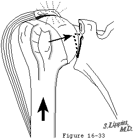
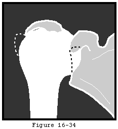
The direction of the net humeral joint reaction force is controlled actively by the elements of the rotator cuff and other shoulder muscles (see figure 33). Neural control of the magnitude of the different muscle forces provides the mechanism by which the direction of the net humeral joint reaction force is modulated. For example by increasing the force of contraction of a muscle whose force direction is parallel to the glenoid center line the body can change the direction of the net humeral joint reaction force to an orientation of closer alignment with the glenoid fossa (see figure 34).
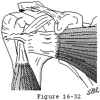
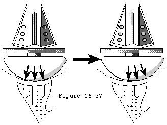
In glenohumeral arthritis control of the net humeral joint reaction force may be compromised by tendon ruptures tuberosity detachment and by deconditioning (see figure 23). The most striking example is in cuff tear arthropathy where the normally stabilizing cuff muscle forces are compromised (see figures 35-37).
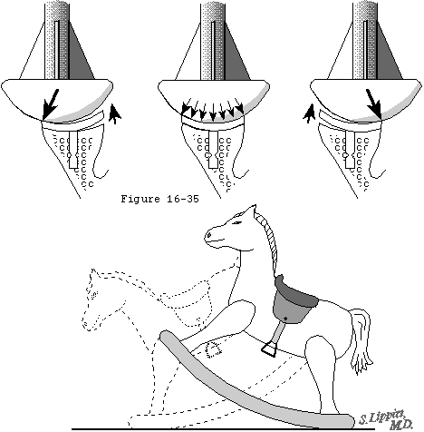
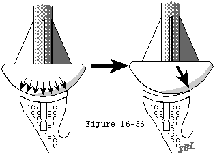
If following glenohumeral arthroplasty the net humeral joint reaction force is not centered in the glenoid fossa eccentric loading may produce rocking horse loosening of the glenoid component (see figure 38). A slight degree of mismatch of the glenoid and humeral diameters of curvature allows for minor amounts of force malalignment before rim contact occurs (see figures 39 and 40). Severt et al (Severt Thomas Tsenter et al. 1993) pointed out that high degrees of conformity between the glenoid and humeral joint surfaces increases the translational forces and frictional torque applied to the glenoid component and on this basis advocated the use of less conforming and less constrained designs.
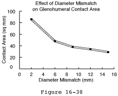
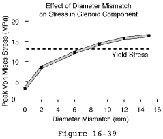
Severe degrees of mismatch may have adverse effects on the glenohumeral contact area (see figure 41) and peak stresses in the polyethylene (see figure 42).
The requisites of strength include:
a. A functional deltoid.
b. A functional rotator cuff.
c. Normal length relationships of muscle origin and insertions.
Surgery and stuffing
In the arthritic shoulder strength can be compromised by cuff deterioration disuse previous injury and previous surgery. The surgeon may be able to enhance the strength of the shoulder through muscle balancing tendon repairs tuberosity reattachment and effective rehabilitation. (Brems 1994) It is critical that the procedure not impair the function of the muscle-tendon units (see figure 43).
The amount of stuffing of the joint sets the resting length of the cuff muscles and to a lesser extent that of the deltoid. If the components are too small the cuff will be slack at rest and thus place the muscles at the low end of the ideal length-tension relationship. If the joint is overstuffed the cuff muscles may be at the high end of their length-tension curve. The distance between the effective cuff insertion and the humeral head center establishes the moment arm for the cuff.
Jacobson and Mallon have provided a method for measuring the glenohumeral offset ratio (Jacobson and Mallon 1993) while Hsu et al have reviewed the influence of abductor lever arm changes after shoulder arthroplasty. (Hsu Wu Chen et al. 1993)
The anatomic requisites of smooth motion are:
a. Smooth joint surfaces
In the normal shoulder intact articular cartilage covering the humeral head and glenoid lubricated with normal joint fluid provide the lowest possible resistance to motion at the joint surface. In arthritis these factors are compromised. Although prosthetic joint surfaces offer much less friction than bone rubbing on bone they have a coefficient of friction approximately ten times greater than that of normal cartilage moving on normal cartilage.
b. Humeroscapular motion interface
A smooth and unimpaired humeroscapular motion interface. The proximal humerus and rotator cuff must slide smoothly beneath the deltoid acromion coracoacromial ligament coracoid and coracoid muscles (see figure 44). Smoothness of the humeroscapular motion interface is often compromised in post surgical and posttraumtic arthritis. The surfaces of this important interface must glide smoothly on each other at the conclusion of the arthroplasty procedure.


