Last Updated: Monday, February 4, 2013
Diagnosing problems of shoulder roughness
In order for the shoulder to function effectively smooth movement must occur in a number of critical joints and motion interfaces. These include the joints:
- between the humerus and scapula (glenohumeral or "ball and socket" joint)
- between the clavicle and the scapula (acromioclavicular joint) and
- between the clavicle and the sternum (sternoclavicular joint).
Smooth motion must also occur at the motion interfaces:
- between the shoulder blade and the chest wall (scapulothoracic motion interface) and
- between the upper arm and the surrounding tissues (humeroscapular motion interface).
Roughness catching grinding crunching or snapping at any of these locations may interfere with the functioning of the shoulder.
Just because noise is produced on moving the shoulder doesn't mean that a serious problem is present. On the other hand loss of the smooth motion of the shoulder can deprive the joint of its normal comfort range of motion and function.
A good history and physical examination along with quality plain X-rays provide sufficient information to diagnose most of the important problems of shoulder roughness.
Click to play

Evaluation of the
Rough Shoulder:
Joints and
Motion Interfaces
There are five areas in which smoothness is required for shoulder function.
Cartilage articulations
Three of these are cartilage to cartilage articulations: the glenohumeral acromioclavicular and sternoclavicular joints. These joints are stabilized by joint capsules ligaments and intraarticular labra or menisci. The smoothness of their cartilage surfaces is at risk for congenital metabolic traumatic degenerative septic and non septic inflammatory joint disease. By selecting the links in the paragraphs below you can see some of the necessary and sufficient criteria we use in making different diagnoses of shoulder roughness.
Five areas
Collapse of the bone supporting the joint surface may be caused by avascular necrosis tumor or osteomyelitis. Labral tears or loose bodies may become interposed between the articular surfaces causing joint roughness.
At the glenohumeral joint different processes produce different patterns of joint surface destruction.
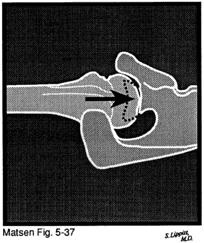
In degenerative joint disease the glenoid cartilage and subchondral bone are typically worn posteriorly sometimes leaving intact articular cartilage anteriorly. The cartilage of the humeral head is eroded in a "Friar Tuck" pattern of central baldness often surrounded by a rim of remaining cartilage and osteophytes. If similar findings arise after an injury or other cause the condition is called secondary degenerative joint disease.
In inflammatory arthritis such as rheumatoid arthritis of the shoulder the cartilage is usually destroyed evenly across the humeral and glenoid joint surfaces.
Cuff tear arthropathy occurs when a chronic large rotator cuff defect subjects the uncovered humeral articular cartilage to abrasion by the undersurface of the coracoacromial arch. The erosion of the humeral articular cartilage begins superiorly rather than centrally. Neurotrophic arthropathy arises in association with syringomyelia diabetes or other causes of joint denervation. The joint and subchondral bone are destroyed because of the loss of the trophic and protective effects of its nerve supply.
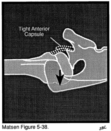
In capsulorrhaphy arthropathy prior surgery for glenohumeral instability leads to joint surface destruction. In this situation excessive anterior or posterior capsulorrhaphy produces obligate translation which forces the head of the humerus out of its normal concentric relationship with the glenoid fossa. The eccentric glenohumeral contact increases contact pressures and joint surface wear. Most commonly over tightening of the anterior capsule produces obligate posterior translation posterior glenoid wear and central wear of the humeral articular cartilage.
The other two locations requiring smoothness are atypical articulations: the scapulothoracic motion interface and the nonarticular humeroscapular motion interface. In these locations motion occurs between tissue planes rather than at joints lined with articular cartilage.
Malalignment of the sliding surfaces surface irregularities or thickening of interposed tissue can interfere with smooth motion at these articulations. One of the more common of these clinical conditions is subacromial abrasion.
Smoothness and motion
The concepts of smoothness and motion are closely related. If the glenohumeral joint surfaces are rough because of degenerative glenohumeral joint disease for example the shoulder will have a marked tendency to become stiff. Restoration of function to such a joint may require not only a resurfacing arthroplasty to restore glenohumeral smoothness but also a capsular release and tendon lengthening to restore motion. Yet lack of smoothness and stiffness need not coexist. Avascular necrosis with collapse of subchondral bone deprives the shoulder of normal smoothness but is not usually associated with stiffness. Conversely a frozen shoulder deprives a shoulder of its motion yet joint surface roughness is not present. Because these two parameters of normal joint function are distinguishable and require separate and distinct treatment we discuss them in two different sections.
History
The history includes a description of the onset of the problem the mechanism of any injuries and the nature and progression of functional difficulties. Systemic or polyarticular manifestations of sepsis degenerative joint disease or rheumatoid arthritis may provide helpful clues. A past history of steroid medication fracture or working at depths may suggest the diagnosis of avascular necrosis. Past injury or surgery increases the risk of infection scarring or abnormal surface contours. Disuse may give rise to abnormal relative positions of the moving surfaces.
The symptoms from lack of smoothness typically occur during use of the shoulder. Often the patient can describe certain motions that are problematic or specific maneuvers that are required to "unlock" or get past a certain sticking point. Occasionally patients will describe a sensation of apparent instability or unwanted shifting of the shoulder. The positions and circumstances which elicit the functional problem must be carefully defined in the history. The patient should also be asked about the response of the shoulder to previous treatment including exercises injections physical therapy and surgery.
The age of the patient at the time of presentation may provide valuable clues to the diagnosis. We have some data on the age at the time of presentation for: degenerative joint disease (see figure 1) rheumatoid arthritis (figure 2) capsulorraphy arthropathy (figure 3) avascular necrosis (figure 4) and cuff tear arthropathy (figure 5).
The physical examination
Physical examination includes the careful observation of the patient's posture for asymmetrical shoulder drooping and muscle atrophy. The rhythm of active rotation and elevation in different planes is observed for breaks in continuity. The patient is asked to demonstrate any maneuvers which produce roughness catching snapping or locking and to localize the site of the problem by pointing with the opposite finger. Patients are usually quite able to indicate one of the five anatomic sites commonly associated with roughness.
The examiner can help distinguish scapulothoracic roughness from glenohumeral problems or from problems at the humerothoracic motion interface by selectively restricting the motion at first one site and then the other. Shrugging protracting and retracting the scapula while the examiner disallows glenohumeral motion permits independent assessment of the smoothness of the scapulothoracic motion interface. Palpation for the site of roughness may localize the problem to the superior medial border of the spine of the scapula. Alternatively rotating and elevating the arm while the examiner stabilizes the clavicle acromion and scapular spine on the chest wall allows independent evaluation of the glenohumeral joint and the humeroscapular motion interface. Roughness in the subacromial area of the nonarticular humeroscapular motion interface is usually manifested on rotation of the arm near 90 degrees of humerothoracic elevation a position in which the capsule is normally lax. Crepitance on this maneuver which reproduces the patient's complaint constitutes a positive subacromial "abrasion sign." Roughness between the subscapularis insertion and the short head of the biceps is evident on rotation of the arm at the side while the biceps is isometrically tightened. Crepitance at the glenohumeral joint is often best palpated posteriorly just beneath the angle of the acromion. It may be accentuated by pressing the humerus toward the glenoid while the joint is rotated. Symptoms from the sternoclavicular or acromioclavicular joints are usually easy to localize on physical examination.
The tenor as well as the location of the noise gives a clue to its etiology. For example a snapping scapula usually produces a low-pitched clunking similar to the noise produced when two sets of knuckles are rubbed against each other. Subacromial abrasion usually produces a higher-pitched crepitance like the sound of wadding up a piece of paper. Dry bone-on-bone grating is typical of roughness of the glenohumeral articular cartilage producing a grating like sandpaper on wood.
Because shoulder roughness may be accompanied by shoulder stiffness and weakness the range of glenohumeral and scapulothoracic motion and the strength of the shoulder motors should be recorded.
Radiographs
The history and physical examination should point to the likely cause and the functional significance of the roughness. The clinical examination will suggest which radiographs may be helpful. Thus the radiographic evaluation is customized to the patient's clinical presentation rather than ordered as part of a "routine."
Scapulothoracic roughness should be evaluated by an anteroposterior view in the plane of the scapula (see figure 6) and by a lateral view in the plane of the scapula (see figure 7) to reveal osteochondromata or malunited fractures of the scapula or ribs. Computerized tomography (CT) can help localize the sites of specific entities but is of minimal value in evaluating a snapping scapula resulting from abnormal posture.
A coned down view of the acromioclavicular joint and an axillary radiograph provide a good two-plane evaluation of this articulation.
Sternoclavicular roughness can be best evaluated with a CT scan.
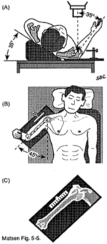
The glenohumeral joint is radiographed using an anteroposterior view in the plane of the scapula and a true axillary view. If the arm is placed in the "centered position" the middle of the humeral articular surface is in the middle of the glenoid fossa (see figure 8). An anteroposterior view and an axillary view taken with the arm in this centered position provide excellent opportunities to evaluate the thickness of the cartilage space between the subchondral bone of the humerus and that of the glenoid to assess the regularity of the subchondral bone and to evaluate any translation of the head of the humerus relative to the glenoid. The anteroposterior radiograph taken in the scapular plane with the arm in the centered position places the humeral neck in maximal profile which is required for accurate use of a humeral prosthesis template.
Click to enlarge

Figure 1 - Age at the time
of presentation for
degenerative joint disease
Click to enlarge

Figure 2 - Age at the time
of presentation for
rheumatoid arthritis
Click to enlarge

Figure 3 - Age at the time
of presentation for
capsulorraphy arthropathy
Click to enlarge

Figure 4 - Age at the time of
presentation for avascular
necrosisavascular necrosis
Click to enlarge

Figure 5 - Age at the time
of presentation for
cuff tear arthropathy
Click to enlarge
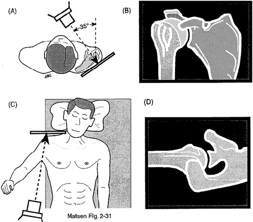
Figure 6 - Anteroposterior
view in the plane of the scapula
Click to enlarge
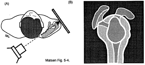
Figure 7 - Lateral view
in the plane of the scapula
Click to enlarge
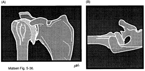
Figure 9 - Radiographs
of dejenerative joint disease
Fortuitously the anatomy of the proximal humerus and the relationship of the scapula on the chest wall make it possible to obtain radiographs which reveal simultaneously the profile of the proximal humerus and glenoid. Because this view centers the head of the humerus on the glenoid it also is the projection most likely to reveal the thinning of the central aspect of the humeral articular cartilage typical of degenerative joint disease (the "Friar Tuck" pattern) whereas radiographs with the arm in other positions may indicate the presence of a thicker layer of cartilage at the periphery of the head.
The relevant anatomy is straightforward. The plane of the scapula makes a 35 degree angle with the plane of the thorax. The humeral neck is in 35 degrees of retroversion with respect to the forearm of the flexed elbow. The humeral neck is also at 45 degrees with the long axis of the humeral shaft. Thus if the forearm of the flexed elbow is perpendicular to the plane of the thorax and if the humerus is abducted 45 degrees the center of the humeral head is pointed at the center of the glenoid. With the arm in this position an anteroposterior radiograph in the plane of the scapula will reveal the desired relationships (see figure 8).
In degenerative joint disease these radiographs (see figure 9) typically show narrowing of the cartilage space between the humeral head and the glenoid sclerosis osteophyte formation and a posterior wear pattern in which the humeral head is posteriorly subluxated in association with erosion of the posterior half of the glenoid. This posterior subluxation may be particularly marked in capsulorraphy arthropathy (see figure 10). In avascular necrosis the predominant radiographic finding is collapse of the subchondral bone of the head of the humerus. In advanced rheumatoid arthritis (see figure 11) the predominant findings usually include loss of the cartilage space between the humerus and the glenoid erosions at the margins of the humeral articular surfaces medial erosion of the glenoid and generalized osteopenia; these changes are often symmetrical affecting both glenohumeral joints.
The bony anatomy of the humeroscapular motion interface can be seen on the anteroposterior view in the plane of the scapula the lateral view of the scapula and the axillary view. These radiographs may reveal a narrowed radiographic acromiohumeral interval sclerosis of the undersurface of the acromion acromial anomalies traction spurs in the coracoacromial ligament and malunited or nonunited fractures of the acromion. These views may demonstrate other potential causes of roughness in the nonarticular humeroscapular motion interface such as anomalies of the proximal humerus malunited tuberosity fractures and functionally significant calcium deposits in the cuff tendons. We have not found the shape of the acromion itself to be useful for separating those shoulders having subacromial roughness from those which do not.
Imaging of the rotator cuff is only carried out if it will affect management of the patient. If the patient meets our criteria for exploration of the subacromial space as described below we will usually avoid cuff imaging because we will be able to evaluate the cuff directly at surgery and will have obtained preoperatively the patient's permission to perform any indicated cuff surgery.
Quality of life
Using the Simple Shoulder Test we collected data on the functional effects of some common causes of shoulder roughness when patients presented for evaluation. We have data for degenerative joint disease (figure 12) rheumatoid arthritis (figure 13) capsulorraphy arthropathy (figure 14) avascular necrosis (figure 15) and cuff tear arthropathy (figure 16).
We have also completed an extensive study of "Patient Self-Assessment of Health Status and Function in Glenohumeral Degenerative Joint Disease" which we present here.
Orthopedists are vitally concerned with optimizing the quality of life for their patients. The quantification of health status and function is central to understanding the impact of chronic musculoskeletal conditions and to determining the effectiveness of different management strategies. With the growing interest in managing health and health care such measurements may help determine which conditions and which treatments merit the highest priority.
Recently patient self-assessment questionnaires have been established as meaningful and practical tools for evaluating health status and function. The effects of musculoskeletal conditions are often quite apparent to the patient; thus these effects are readily detectable by patient self-assessment.
The purposes of this article are:
- to demonstrate the practicality of office-based patient self-assessment in the documentation of health status and function in a population of individuals with a well-defined musculoskeletal condition: primary glenohumeral degenerative joint disease
- to compare the health status results with those expected in a general population that is age matched and
- to determine which general health status parameters were most closely associated with loss of shoulder function.
Methods: Patient population
This study concerns 103 consecutive patients presenting to the senior author for evaluation and management of primary glenohumeral degenerative joint disease. Each patient met established necessary and sufficient conditions for this condition. Seventy-seven were male twenty-six female. The mean age was sixty-three years (± 13 SD range 30-94). Sixty-three were right dominant thirty-eight were left non-dominant seven were left dominant and five were right non-dominant.
Self-assessment of health status
Each patient completed a questionnaire consisting of thirty-six questions regarding their general health status the Short Form-36 (SF 36). The health status questions were scored using an established protocol and converted into eight health status parameter scores for each of which "100" represented the most healthy and "0" the least healthy score. The data from the 103 subjects with primary glenohumeral degenerative joint disease were compared to the published results using the same health status questionnaire for three separate population-based health status surveys: the Geisinger Health Plan Survey (1 760 subjects) the AT&T American Trans Tech "MASH" Trial (702 subjects) and the Northwest Area Foundation Health Survey (1 814 subjects). Initially the comparison was made for men and women separately but the sex-related differences were small and these have been omitted from this presentation for reasons of brevity. The reference data cohorts did not exclude patients with comorbidities. For those subjects under sixty-five years of age the most prevalent chronic diseases included chronic low back pain (11.1%) arthritis (9.6%) asthma hypertension and visual impairment. Among the subjects over the age of sixty-five the most prevalent conditions were arthritis (56.3%) chronic low back pain (37.5%) hypertension angina and gastrointestinal problems. Thus the referenced data represent a cross-section of the populations studied and do not represent the health status of disease-free individuals.
The health status scores of the SF 36 are age dependent; thus both the data on our patients and the reference cohort data were graphed as a function of patient age. For each health status parameter the means means plus one standard deviation and the means minus one standard deviation for the combined reference cohort were plotted. For each health status parameter the percent of patients more than one standard deviation below the mean were determined from these graphs.
Self-assessment of shoulder function
Each patient completed twelve questions concerning the function of their shoulder the Simple Shoulder Test (SST ). Comparison shoulder function data were not available on the same population used for the health status reference. Instead we compared the shoulder function of our patients to that of 80 individuals aged 60-70 years who had no evident shoulder disease on a standardized history physical and ultrasonographic examination of the rotator cuff. Of these 80 patients all could perform all twelve of the simple shoulder test functions except for one patient who could not lift eight pounds to shoulder level and three who could not throw overhand twenty yards.
Results: Self-assessment of health status
We prepared plots of pain (see figure 17) and physical role function (see figure 18) scores for each of the 103 subjects as a function of the patient's age. (In these plots the dots indicate subjects from this study. Lines demonstrate mean ± standard deviation data from population-based comparison cohort). Similar plots were carried out for the six other health status parameters. On each of these graphs the number of subjects scoring more than one standard deviation below the mean was counted and expressed as a percent of the total number of patients. We determined the percent of patients who were more than one standard deviation below the mean for each of the eight health status parameters (see figure 19). (In this plot if all distributions were normal seventeen percent of the subjects would have been expected to lie more than one standard deviation below the population-based mean (vertical line)). For example over 50% of the patients' pain and physical role functioning scores were more than one standard deviation below the mean. If the distribution of the two populations had been normal only 17% of the subjects would score more than one standard deviation below the mean.
Shoulder function
A substantial number of subjects were unable to perform each of the twelve shoulder functions (see figure 20). Over 50% of subjects were unable to sleep on the affected side wash the back of the opposite shoulder place their hand behind their head with the elbow out to the side reach their low back to tuck in a shirt and toss twenty yards overhand.
Discussion
This study demonstrated that both the quality of life and the shoulder function were compromised in this series of 103 patients with primary glenohumeral degenerative joint disease. These patients are obviously a subset of patients meeting the criteria for this diagnosis: they were sufficiently impaired to present to our referral medical center for evaluation and management of their disease. Thus these results may not be representative of the population of patients with primary glenohumeral degenerative joint disease or those presenting in other practice settings.
While this is one of the first studies to apply the method to shoulder disease the use of self-assessment tools to document the impact of musculoskeletal conditions has been recently demonstrated by others. These studies indicate that musculoskeletal conditions when compared to other medical disorders have a great impact on health and function. In this study most of the health status parameters derived from the SF 36 were lower in these patients with primary glenohumeral degenerative joint disease than for general comparison populations. This is of interest because none of the health status parameters of the SF 36 directly assess upper extremity function.
While many orthopedic scoring systems have been developed to document disease severity many of these scoring systems focus on "objective" parameters such as range of motion strength and radiographic appearance. The SF 36 and other self-assessment instruments have the advantage of emphasizing the patients' perspective. Self-assessment forms are also more practical (less patient time less cost) to administer and offer the potential for periodic followup assessments without the patient having to return to the office.
Short form generic health surveys such as the SF 36 have been shown to be as effective and reliable as the longer surveys. The SF 36 has also been shown to be useful in documenting the outcome of orthopedic surgery. The importance of the SF 36 to orthopedics is that this instrument is used in other fields of medicine as well; thus the impact of musculoskeletal problems on self-assessed health status can be compared to the impact of other chronic conditions such as endometriosis renal failure angina gastrointestinal disease and hypertension. The generality of the SF 36 also means that conditions other that the one under study (comorbidities) may affect the results. The published reference health status parameter data indicate a trend for diminished scores with increasing age no doubt reflecting a growing prevalence of comorbidities with age. In comparison to the reference populations the distribution of bodily pain and physical role function scores for the subjects with primary glenohumeral degenerative joint disease were skewed so that over 50% of the subjects were more than one standard of deviation below the referenced mean.
For the study of shoulder disease the Simple Shoulder Test provides a needed compliment to the SF 36. In performing the twelve functions of the SST subjects have been shown to use the shoulder in a wide variety of positions ranging from sixty degrees of elevation in the minus fifty degree thoracic plane (tucking in the shirt) to 120 degrees of elevation near the coronal plane (placing the hand behind the head with the elbow out to the side) to seventy degrees of elevation in the plus 130 degree thoracic plane (washing the back of the opposite shoulder). As a group the patients with primary glenohumeral degenerative joint disease had much poorer shoulder function than the nearly perfect function of apparently disease-free shoulders of similar age.
Some of the health status parameters correlated strongly with the patients' ability to perform different shoulder functions. Overall bodily pain and physical functioning were the most strongly affected. In the future study of the effectiveness of treatment of shoulder disorders will indicate whether improvements in these health status parameters parallel improvements in the shoulder functions.
Click to enlarge

Figure 12 - Functional
deficits of patients with
degenerative joint disease
Click to enlarge

Figure 13 - Functional
deficits of patients
with rheumatoid arthritis
Click to enlarge

Figure 14 - Functional
deficits of patients with
capsulorraphy arthropathyapsulorraphy
arthropathy
Click to enlarge

Figure 15 - Functional deficits
of patients
with avascular necrosis
Click to enlarge

Figure 16 - Functional deficits
of patients with
cuff tear arthropathy
The SF 36 and SST represent practical examples for generic and condition-specific measurement of the health and functional status in patients with primary glenohumeral degenerative joint disease. Our subjects had no difficulty in completing these self-assessment questionnaires. The collection of these data did not require physician or staff time other than passing out and collecting the forms. The Simple Shoulder Test requires no calculation. The standardized algorithms for calculating the SF 36 health status parameters are easily incorporated into a spreadsheet. No research person or specialized equipment was required to collect or analyze these data. The incorporation of these tools into the context of a busy office practice provides a practical method for quantitating the impact of shoulder conditions on health status and shoulder function.
