Last Updated: January 26, 2005
About cuff defects
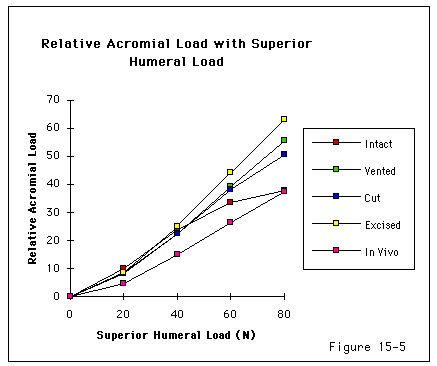
The requisites for normal cuff function are stringent including healthy strong cuff muscles, normal capsular laxity intact cuff tendons, a smooth contour of the undersurface of the coracoacromial arch, a thin lubricating bursa, a smooth upper surface of the cuff and tuberosities and concentricity of the glenohumeral and cuff-coracoacromial spheres of rotation (see figures 1-5). Disorders of this complex mechanism constitute the most common source of shoulder problems. (Chakravarty and Webley 1993 Iannotti 1994)
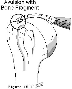
Cuff disruption may be partial or full thickness acute or chronic and traumatic or degenerative. (Cofield 1985 Matsen Lippitt 1994) The magnitude of cuff disruption ranges from the mildest strain to total absence of the cuff tendons. In younger patients partial thickness cuff lesions may include the avulsion of a small chip of bone from the tuberosity the radiographic appearance of which should not be confused with that of calcific tendinitis (see figure 6). Contributing factors may include trauma (Codman 1911 Codman 1937) attrition (DePalma Gallery 1949 Keyes 1935 Meyer 1924 Moseley 1952) ischemia (Lindblom 1939a Lindblom and Palmer 1939 Moseley and Goldie 1963 Rathbun and Macnab 1970 Rothman and Parke 1965) and subacromial abrasion. (Craig 1984 Neer 1972 Neer 1983 Neviaser and Neviaser 1982 Peterson and Gentz 1983 Watson 1978)
Degenerative cuff failure almost always starts with a partial thickness defect on the deep surface near the attachment of the supraspinatus to the greater tuberosity. Codman's view of the frequency of this lesion and the potential range of pathology is indicated by the following passage (Codman 1934b)
Figure 7 shows an extensive tear so that the rent has come through to the most superficial fibers of the tendon. The reader should visualize this vertical section so as to understand that the rent also extends along the curve of the edge of the joint cartilage to a considerable extent leaving the sulcus bare perhaps for an inch or more. This condition I like to call a "rim rent and I am confident that these rim rents account for the great majority of sore shoulders. It is my unproved opinion that many of these lesions never heal, although the symptoms caused by them usually disappear after a few months. Otherwise, how could we account for their frequent presence at autopsy?
The anatomically observed prevalence of partial thickness cuff lesions leads one to Codman's suggestion that commonly-diagnosed diagnoses of shoulder pain, referred to as cuff tendinitis" "bursitis" or "impingement syndrome" may actually represent failure of the deep surface fibers of the rotator cuff. (Fukuda Hamada 1994) The degree to which the fibers that remain intact may hypertrophy strengthen or adapt (Burkhart Fischer 1996) to stabilize the tear and take up the function of the damaged fibers are not known. It appears likely that repeated failure of small groups of fibers leads not only to self-limited acute symptoms (perhaps interpreted as "tendinitis or bursitis" [Hawkins Misamore 1985]) but also to progressive weakness of the rotator cuff making it increasingly susceptible to damage from lesser loads. This gives rise to the "creeping tendon ruptures" described by Pettersson. (Pettersson 1942) The observation by Pettersson (Pettersson 1942) and others that major cuff defects may occur without symptoms or recognized injury suggests that previous minor often subclinical fiber failure leaves shoulder weaker and the cuff tendons progressively less able to withstand the loads encountered in daily living.
The incidence of rotator cuff tendon defects has been described in various reports of cadaver dissections: Smith (Smith 1834) found an incidence of 18 per cent; Keyes (Keyes 1933) 19 per cent; Wilson (Wilson 1943 Wilson and Duff 1943) 20 per cent in a series of autopsy dissections and 26.5 per cent in a series of cadaver dissections; Cotton and Rideout (Cotton and Rideout 1964) 8 per cent; Yamanaka and coworkers (Yamanaka Fukuda 1983) 7 per cent; Fukuda and associates (Fukuda Mikasa 1987) 7 per cent; and Uhthoff and colleagues (Uhthoff Loehr 1986 Oct 27) 20 per cent.
More about epidemiology
Neer found that the incidence of complete cuff tears in more than 500 cadaver shoulders was less than 5 per cent. (Neer 1983) Lehman et al (Lehman Cuomo 1995) found that the incidence of full thickness rotator cuff tears in 235 male and female cadavers ranging in age from 27-102 years (average 64.7 years) was 17% (53 female 26 male). The average age of those cadavers with tears was 77.8 years as compared to 64.7 years in the intact group. Recognizing the importance of age in the prevalence of cuff lesions these authors noted that in cadavers under 60 years of age the incidence of rotator cuff tears was 6% as opposed to 30% in those over 60 years of age.
Partial-thickness tears appear to be about twice as common as full thickness defects. Yamanaka and coworkers (Yamanaka Fukuda 1983) and Fukuda (Fukuda 1980 Fukuda Mikasa 1983 Fukuda Mikasa 1987) reported on 249 cadaver left shoulders in which they found a 13 per cent incidence of partial-thickness tears. Thirty per cent of shoulders over 40 had cuff tears whereas there were no tears seen in those under 40. Three percent had tears on the bursal side three percent had tears on the joint side and 7 percent had intratendinous tears. In another clinical series of partial-thickness cuff tears Fukuda and associates (Fukuda Mikasa 1983) found 9 tears on the bursal side 11 on the joint side and 1 intratendinous. The bursal side tears had the most severe symptoms. All of these tears were localized in the critical area of the supraspinatus tendon. In his studies of 96 shoulders in patients ranging in age from 18 to 74 years DePalma found a 37 per cent incidence of partial-thickness tears of the supraspinatus and infraspinatus a 21 per cent incidence of partial-thickness tears in the subscapularis and a 9 per cent incidence of full-thickness tears. Uhthoff and associates(Uhthoff Loehr 1986 Oct 27) found a 32 per cent incidence of partial-thickness tears in 306 autopsy cases with a mean age of 59 years. Other studies report partial-thickness tears in approximately 20 to 30 per cent of cadaver shoulders. (See references Codman 1937 Cofield 1985 Cotton and Rideout 1964 Fukuda Mikasa 1983 Grant and Smith 1948 Hawkins Misamore 1985 Keyes 1933 Lindblom 1939a Lindblom 1939b Lindblom and Palmer 1939 Uhthoff Loehr 1986 Oct 27) The data from studies in which the cuff was sectioned to demonstrate the prevalence of intrasubstance lesions indicate that cadaver or clinical examinations confined to the bursal and articular sides of the tendon will overlook the common intratendinous form of cuff defect.
The incidence of cuff defects in living subjects is more difficult to study. In a community survey of 644 individuals over 70 years of age Chard et al (Chard Hazleman 1991) found 21% had shoulder symptoms (25% in women 17% in women) the majority of which were attributed to the rotator cuff. However fewer than 40% of these subjects sought medical attention for these symptoms.
Distorted views of the incidence of cuff disease and of the relationship of cuff tears to clinical symptoms are obtained if only symptomatic patients are studied. Thus some of the most important studies have concerned the prevalence of cuff lesions in asymptomatic patients. Pettersson (Pettersson 1942) performed arthrography on 71 apparently healthy asymptomatic shoulders ranging in age from 15 to 85 years. He found that of 27 asymptomatic untraumatized shoulders in patients aged 55 to 85 13 had arthrographically proven partial- or full-thickness rotator cuff defects most were observed between the ages of 70 and 75 years. All these shoulders were symptom free and without history of trauma. Repeated episodes of fiber failure lead to progressive cuff weakness but not necessarily to pain unless the extension of the defect is acute and substantial. Milgrom et al (Milgrom Schaffler 1995) found that the prevalence of partial- or full-thickness tears increased markedly after 50 years of age: over 50% of subjects in their seventh decade and over 80% in subjects over 80 years of age. They concluded that "rotator-cuff lesions are a natural correlate of aging and are often present with no clinical symptoms." Sher et al (Sher Uribe 1995) used MRI to evaluate asymptomatic shoulders over a wide age range and found that 15 percent had full thickness tears and 20 percent had partial thickness tears. The frequency of full-thickness and partial-thickness tears increased significantly with age (p < 0.001 and 0.05 respectively). Twenty-five (54 per cent) of the forty-six individuals who were more than sixty years old had a tear of the rotator cuff: thirteen (28 per cent) had a full- thickness tear and twelve (26 per cent) had a partial-thickness tear. Of the twenty-five individuals who were forty to sixty years old one (4 per cent) had a full-thickness tear and six (24 per cent) had a partial-thickness tear. Of the twenty-five individuals who were nineteen to thirty-nine years old none had a full-thickness tear and one (4 per cent) had a partial-thickness tear. They concluded that
- magnetic resonance imaging identified a high prevalence of tears of the rotator cuff in asymptomatic individuals
- these tears were increasingly frequent with advancing age and
- these defects were compatible with normal painless functional activity.
In another most important study Yamanaka and Matsumoto (Yamanaka and Matsumoto 1994) demonstrated the progression of partial thickness tears. After initial arthrography they followed 40 tears (average patient age 61 years) managed without surgery repeating the arthrogram at an average of more than a year later. Although the patients had improved average shoulder scores at followup followup arthrographies revealed apparent resolution of the tear in only four instances reduction of the tear size in only four enlargement of the tear size in 21 and progress to full thickness cuff tear in 11 patients. The authors concluded that tears were likely to progress with increasing age in the absence of history of trauma.
Thus it must be concluded that cuff defects become increasingly common after the age of 40 and that many of these occur without substantial clinical manifestations.
Certain occupations seem to be particularly problematic for the rotator cuff including tree pruning fruit picking nursing grocery clerking longshoring warehousing carpentry and painting. (Luopajarvi Kuorinka 1979) Some patients relate the onset to some type of athletic activity such as throwing tennis skiing and swimming. Richardson and associates (Richardson Jobe 1980) reviewed 137 of the best swimmers in the United States. The incidence of shoulder problems was 42 per cent. These authors calculated that the average national-level swimmer puts his or her shoulder through about 500 000 cycles per season. Although subluxation is a recognized problem in this group many were found to have symptoms and signs suggesting cuff involvement. The technique an athlete uses has a major relationship to the development of or freedom from symptoms as discussed by Richardson and coworkers (Richardson Jobe 1980) Albright and colleagues (Albright Jokl 1978) Cofield and Simonet (Cofield and Simonet 1984) Penny and Welsh (Penny and Welsh 1981) Neer and Welsh (Neer and Welsh 1977) and Penny and Smith. (Penny and Smith 1980)


